Material and method
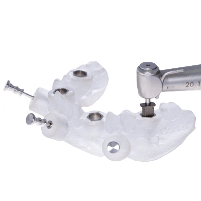
In order to examine the existence of a microgap on dental implants; a special experimental test field was developed. For each type of implant system tested; five inspection pieces were manufactured. Each inspection piece simulates an implant-supported molar crown in the upper jaw. During the load, in a two-dimensional chewing simulator, a constant and diverging X-ray device radiated the inspection pieces.
By transforming of the X-ray into visible light; x-ray videos were recorded, using a high-speed digital camera. The results will give information on the development and a conclusion of a micro gap at the Implant Abutment Interface.
Experimental setup
Illustration 1 shows schematically the experimental setup. Those in the x-ray source: [1] analysation of an exemplary inspection piece loaded in a two-dimensional chewing simulator [3] [2; 4]. The x-rays are converted in the image amplifier [5] into visible light.
The quantity of light coming from the image amplifier meets the CCD sensor of the High-speed digital camera [6]. Afterwards; the camera sends a digital signal to an attached computer, whereas the computer then develops x-ray-videos on the processes of the Implant-Abutment-Interface.
The applied X-ray unit is a constant emitter, with which the X-radiation spreads divertingly. Diversional spreading of the radiation leads to an enlargement of the radiated object (see fig. 3)


